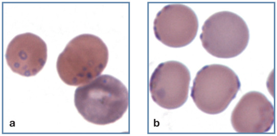
Figure 20.

Large numbers of small blue/purple-staining Mycoplasma haemofelis organisms on the surface of erythrocytes from two cats with a natural infection. (a) Ring forms of the organism can be seen on the surface of an erythrocyte. A polychromatophilic erythrocyte (aggregate reticulocyte) without organisms is also present (bottom right). (b) Organisms appear on the periphery of erythrocytes in a thin area of the blood film. Wright-Giemsa stain
