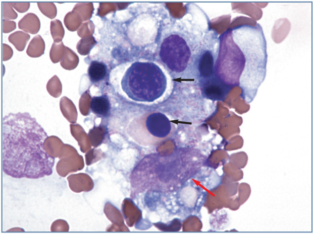
Figure 4.
Aggregate of cells, which appears to contain a macrophage with phagocytized nucleated erythrocytes (black arrows), located at the feathered end of a blood film from a cat with Mycoplasma haemofelis infection. The large oval magenta structure near the bottom of the image (red arrow) is the nucleus of the macrophage. The nucleus contains a blue-staining nucleolus. Wright-Giemsa stain

