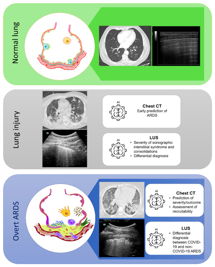
Figure 2.
Current potential areas of application for artificial intelligence applied to lung computed tomography and lung ultrasound imaging in different stages of lung disease. Green upper box: normal lung histology (drawing on the left), axial projection of a lung CT scan (image in the middle), and a LUS scan showing normal pleural findings with repetitive physiological horizontal artifacts (A lines), a typical sign indicating a normal aerated lung. Grey box in the middle: a lung CT scan (upper image) and LUS scan (lower image) in a patient with acute lung injury. Note the presence of vertical artifacts arising from the pleural line (B lines), indicating the presence of a sonographic interstitial syndrome. Blue box at the bottom: overt ARDS (drawing on the left) with alveolar–capillary damage, alveolar edema, cellular debris, neutrophilic migration (in violet), activated macrophages (in yellow), fibroblast activation, and fibrin deposition (in green). The lung CT scan (upper figure in the middle) and the LUS scan (lower figure in the middle) represent the typical radiological findings in a representative patient with ARDS. Note the inhomogeneity of aerated and not aerated parenchyma at the axial projection of the lung CT and the irregular pleural profile, with areas of high lung density (white lung) interspersed by parenchymal subpleural infiltrates. The two different imaging approaches carry different qualitative and quantitative information of the same pathological pattern. AI: artificial intelligence; ARDS: acute respiratory distress syndrome; CT: computed tomography; LUS: lung ultrasound.

