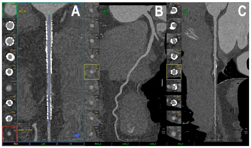
Figure 3.
Cardiac PCCT visualization of coronary stents and stented lumen. There are two stents at the level of the proximal and middle RCA (A) and one stent on the marginal branch of the left LCx (C); the LAD (B) is normal, without any detectable atherosclerotic disease. All stents are perfectly visualized in terms of their inner struts and also in their inner lumen, which is difficult to achieve using standard cardiac CT. PCCT—photon-counting CT, LAD—left anterior descending, LCx—left circumflex, RCA—right coronary artery. Reprinted with permission under open access from Cademartiri et al. [22].

