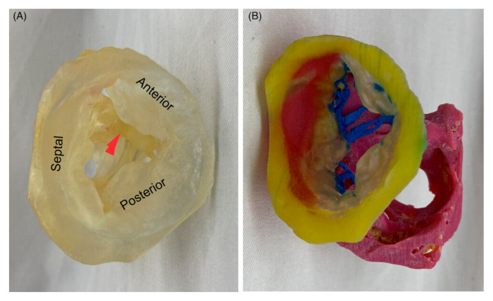
Figure 6.
A 3D-printed model of the tricuspid valve of a human heart specimen (HH 223). (A) A model printed using a clear material as viewed from the atrium, with leaflets labeled and the moderator band marked with a red arrow. (B) A model printed using multiple colors and materials and rotated to show the subvalvular apparatus. Yellow, tricuspid annulus; transparent, mitral leaflets; blue, chordae tendinea; pink, papillary muscles. Reprinted with permission from Arango et al. [103].

