Figure 12.
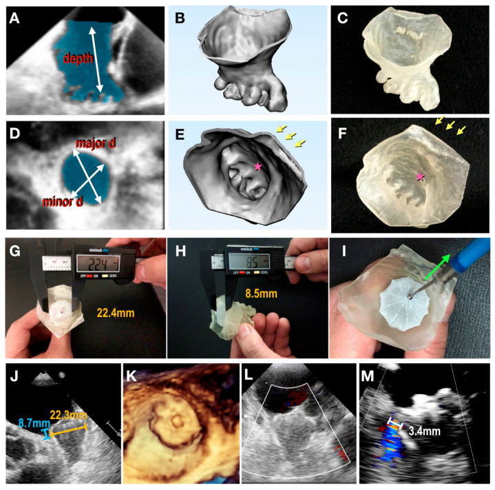
Three-dimensionally printed patient-specific models based on echocardiographic images. (A–F) From 3D transesophageal echocardiography (TEE) image to 3D physical model. (A,D) Segmentation of left atrial appendage (LAA) (shaded area) based on 3D TEE data. Measurements regarding the major and minor ostial diameters and depth of the LAA were taken. (B,E) Creation of a digital object. (C,F) Three-dimensional printed physical model made of tissue-mimicking material. Arrows denote pulmonary vein ridge; stars denote appendicular trabeculations. (G–I) Modifying the size of the 3D model. (G) Device compression and (H) protrusion in 3D model measured using a digital caliper. (I) Tug test for stability. (J) Device compression and protrusion measured in a clinical procedure. (K) Three-dimensional TEE en face view of final device position. (L) Color Doppler assessment showing no peridevice leaks. (M) In another case, color Doppler assessment revealed a residual leak with a jet width of 3.4 mm. Reprinted with permission under open access from Fan et al. [112].

