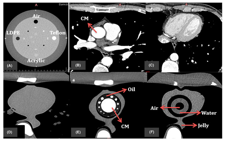
Figure 17.
The resulting axial CT of (A) four inserts in Catphan@ 500 phantom; (B,C) patient image datasets for cardiac CT; (D) original cardiac insert of anthropomorphic chest phantom; (E,F) 3D-printed cardiac insert phantom with the contrast materials (CM), oil, air, water, and jelly segmented all labeled. Reprinted with permission under the open access from Abdullah et al. [139].

