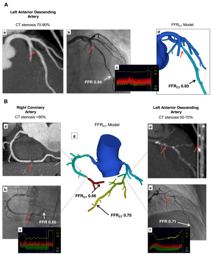
Figure 24.
Examples of FFRCT in assessing the hemodynamic significance of coronary lesions at three main coronary arteries (A,B). Coronary CT angiography shows significant stenoses on the left anterior descending artery (LAD), right coronary artery (RCA), and left circumflex (LCx), while FFRCT shows ischemia at RCA and LCx but not at LAD, as the FFRCT value is more than 0.80. This was confirmed by invasive FFR measurements, as shown in (A(c)) and (B(c,f)). (a,b) in image (A), (a,b,d,e) in image (B) refer to stenotic lesions of RCA and LAD on coronary CT angiography and invasive FFR measurements, respectively, while ((A)d,(B)g) indicate FFRCT measurements at these coronary arteries. Reprinted with permission from Norgaard et al. [43].

