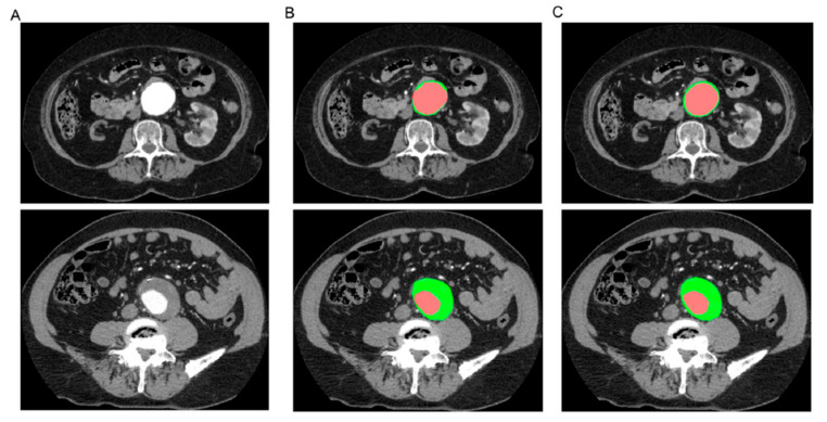
Figure 30.
Representative images of the segmentation of the aortic lumen (in red) and the intraluminal thrombus (in green). (A) CT scan cross-sectional views of patients with infrarenal AAA. (B) Manual segmentation. (C) Automatic segmentation. Reprinted with permission under open access from Lareyre et al. [201].

