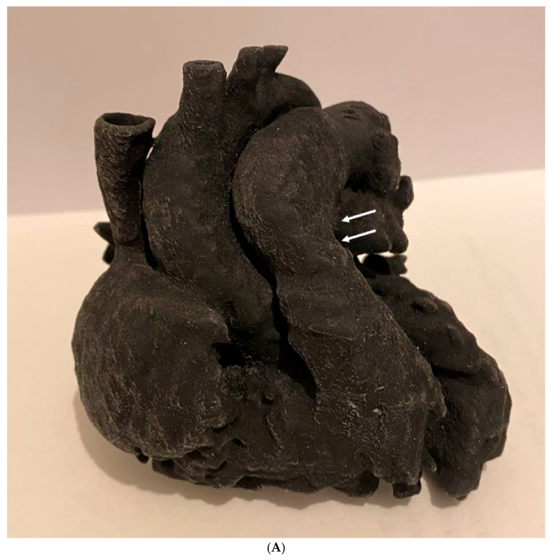
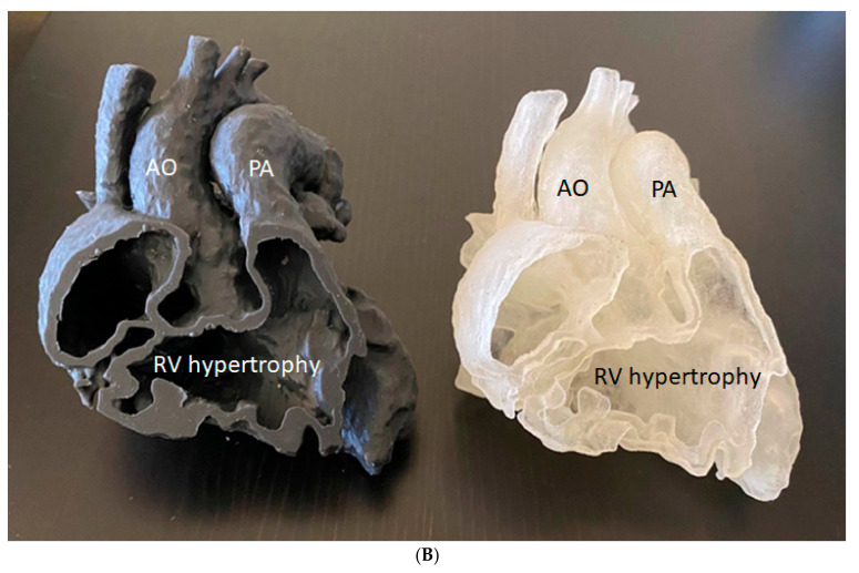
Figure 34.
Three-dimensionally printed heart model of a patient with Tetralogy of Fallot. This model was printed based on cardiac CT images using Agilus30 material, and its tissue properties are similar to those of human heart tissues. The model was printed in one piece (A) and a two halves (B) to show the internal structures. The arrows refer to the pulmonary artery stenoses. AO—aorta, PA—pulmonary artery, RV—right ventricle. Reprinted with permission under open access from Sun [215].


