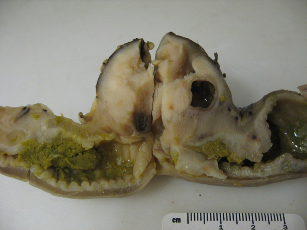
Figure 2.

Gross pathologic cut-section of colonic mass which correlates to the ultrasound image in Figure 1 (cat 2). A focal, well-encapsulated, eccentric mural mass is present within the colon. The well-defined cavitation in the right side of the mass correlates with the circular region containing well-defined areas of hyper- and hypoechogenicity in the ultrasound image (Figure 1), and histologically represents an area of necrosis. There is focal narrowing of the colonic lumen secondary to the mass and there is thickening of the adjacent colonic wall
