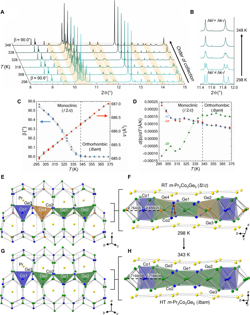
Fig. 1. In situ x-ray diffraction and crystal structure of m-Pr2Co3Ge5 and o-Pr2Co3Ge5.
(A and B) Temperature-dependent x-ray diffraction of m-Pr2Co3Ge5 (λ = 0.458977 Å) highlighting the structural phase transformation from I2/c to Ibam. The change in angle β as related to the change in volume and the change in lattice parameters (lp) are given in (C) and (D), respectively. Dashed lines indicate the conclusion of the second-order structural phase transformation. Crystal structure of room-temperature (E and F) and 343-K (G and H) m-Pr2Co3Ge5 obtained from synchrotron PXRD where Pr, Co, and Ge are represented with yellow, blue, and green/orange spheres, respectively. Select Co-Ge distances (Å) are shown to illustrate the distortion of the basal atoms of the tetrahedral slab along the crystallographic b direction. Red and blue arrows indicate the separation and contraction of the Co2 dimerization, respectively.

