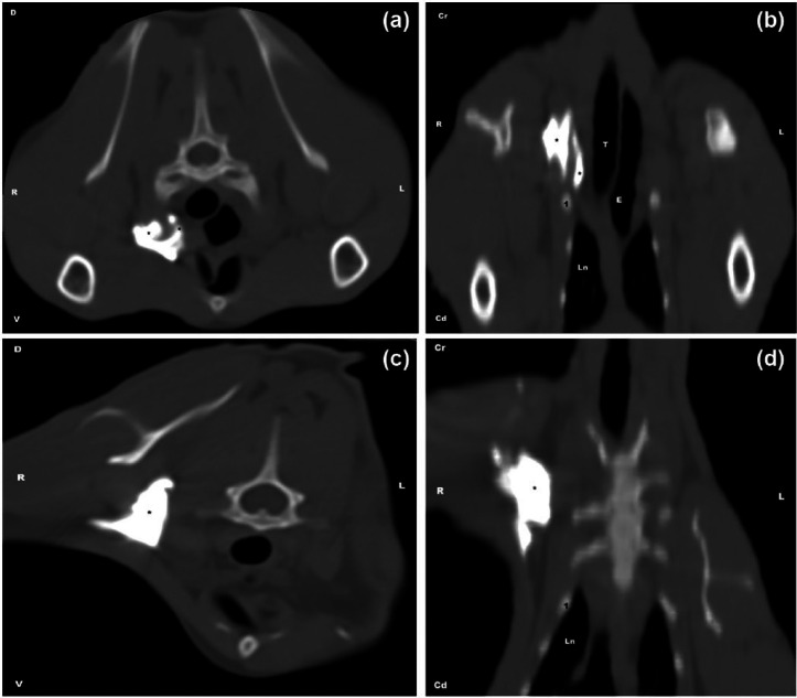
Figure 4.
Pattern of the distribution of the injectate in the CT study. (a) FAD technique: transverse view; the contrast medium (*) is observed spreading at the axillary space and within the thorax. (b) FAD technique: dorsal view. (c) FAB technique: transverse view; the contrast medium (*) is observed spreading at the axillary space. (d) FAB technique: dorsal view.
FAD technique = both forelimbs are adducted; FAB technique = the forelimb to be blocked is abducted 90°; Cr = cranial; Cd = caudal; R = right; L = left; D = dorsal; V = ventral; Ln = lung; 1 = first rib; T = trachea; E = oesophagus

