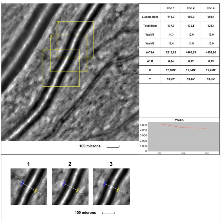
Figure 2.
Image of the supratemporal arteriole in a healthy subject. Evaluation of retinal arteriolar morphology in a POAG patient with adaptive optics camera using 4° × 4° square (Rtx-1, Imagine Eyes, Orsay, France) and measurement of morphological parameters using AOdetect software (AO Image 3.4). The parameters were calculated from the three selected regions of interest (yellow squares) for each time landmark (each with 100 μm width and height) (blue and yellow boxes indicate the walls of the arteriole) (bottom). The chart presents the following parameters: Lumen diam—lumen diameter; Total diam—total diameter; wall1 and wall2; WCSA—cross-sectional area; WLR—wall-to-lumen ratio. The image is from the author’s collection.

