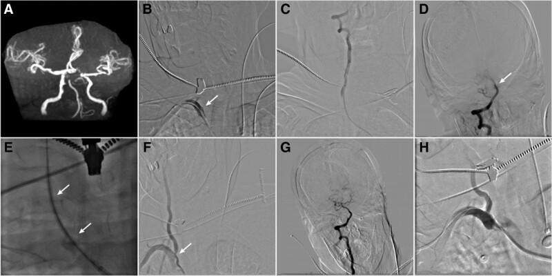
Figure 2.
(Patient 5). (A) cranial MRA showed occlusion of the basilar apex. (B) Right subclavian artery angiography showing the right vertebral artery stump (white arrow). (C and D) 4F MPA catheter angiography showed unobstructed blood flow in the distal vertebral artery with embolization of the basilar artery tip (white arrow). (E) a conical inner dilator passed through the occluded vertebral artery (double white arrow). (F and G) After mechanical thrombectomy, angiography showed stenosis of the right vertebral artery, with a stenosis rate of about 60% (white arrow); the thrombus at the tip of basilar artery had been removed. (H) angioplasty at the ostial of vertebral artery with 4 * 15 mm balloon expandable stent. MRA = magnetic resonance angiography.

