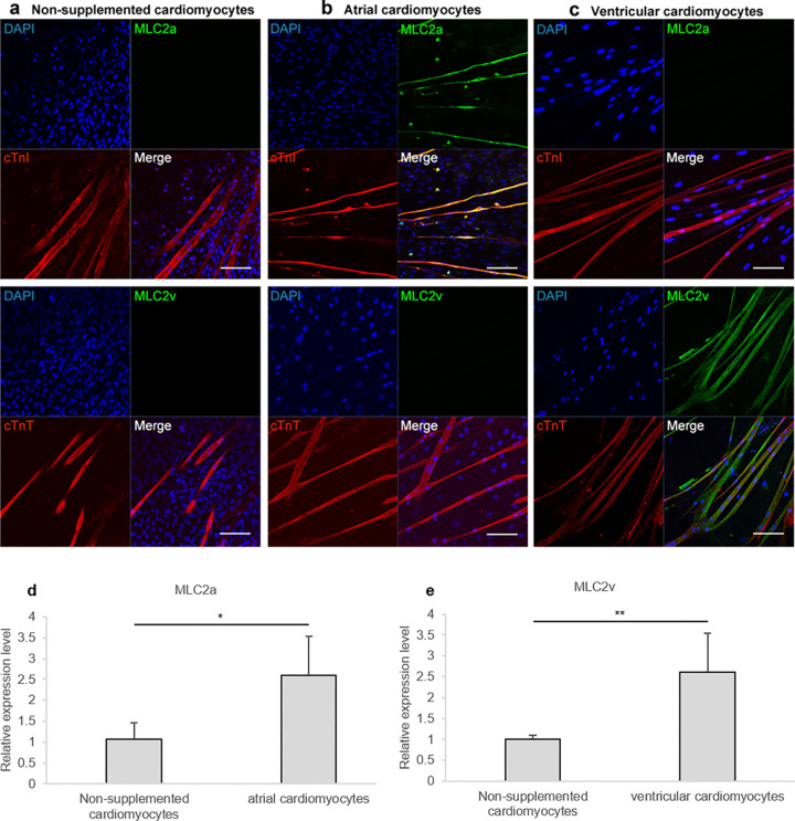
Fig 2. Rat HAP stem cells differentiated into atrial or ventricular cardiomyocytes.
(a-c) Immunofluorescence staining of the upper parts of rat vibrissa hair follicles, which were cultured for 21 days, differentiated into cTnI (a-upper; red) and cTnT (a-lower; red)-positive non-supplemented cardiomyocytes; cTnI (b-upper; red), cTnT (b-lower; red)- and MLC2a (b; green)-positive atrial cardiomyocytes; cTnI (c-upper; red), cTnT (c-lower; red)- and MLC2v (c; green)-positive ventricular cardiomyocytes. Nuclear staining with DAPI (blue). Scale bars; 100 μm. (d, e) qPCR analyses of cardiomyocytes differentiated from HAP stem cells. n = 4 per group for non-supplemented, atrial, and ventricular cardiomyocytes. Data are presented as mean ± SD. * P < 0.05, ** P < 0.005, two-sided Student’s t-test.

