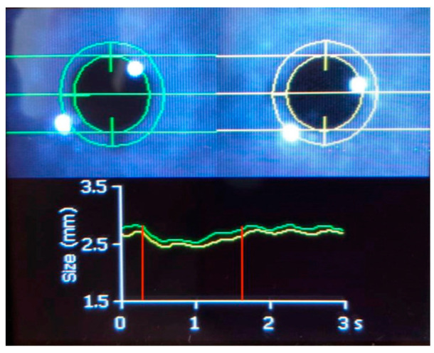
Figure 5.
Automated pupillometry in a 30-year-old woman who was intubated and mechanically ventilated with coma and normal brain imaging. The green line shows the direct pupillary response of the right pupil to a flashlight stimulus and the yellow line shows the response of the left pupil. The pupillary constriction to light (shown in the area between the red vertical lines) is preserved, and an oscillation of approximately 1.5 Hz is superimposed. This phenomenon is hippus [81]. Hippus can be normal or pathological. Hippus comes from the Greek “hippos”, meaning horse, and refers to rhythmic, dilating, and contracting pupillary movements [82]. Hippus has been related to epilepsy; however, in this case, concomitant electroencephalography did not detect epileptic activity.

