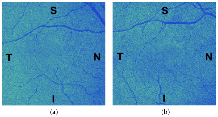
Figure 3.
Example of the mean value fundus (MVF) images computed for the outer nuclear layer of a 54-year-old female participant: (a) the right eye; and (b) the left eye, flipped to match the regions of the right eye. S, T, N, and I stand for the eye’s superior, temporal, nasal, and inferior regions, respectively. These images are shown for reference purposes only; they were intensity-corrected and pseudo-colour-coded for ease of visualisation.

