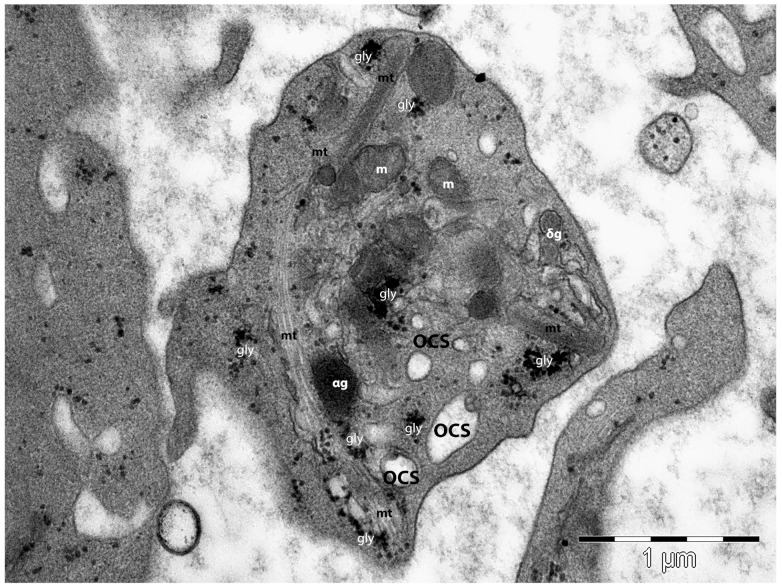
Figure 3.
Transmission electron microscopy image of a platelet. The peripheral distribution of the microtubular (mt) loop is obvious and demonstrates its implication in platelet morphology and shape maintenance. Three-dimensionally open canalicular system (OCS) elements are present, along with glycogen granules (gly), mitochondria (m), α–granules (αg), and δ-granules (δg). (Courtesy of Dr. E.T. Fertig, “Victor Babeş” National Institute of Pathology, Bucharest, Romania).

