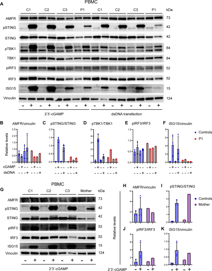
Fig. 2.
Reduced STING signaling and ISG responses in patient cells. A PBMCs from the patient and three healthy controls (C1–C3) were stimulated with 100 µg/mL of 2′3′-cGAMP (A) or 2 µg/mL transfected dsDNA for 3 h and lysates were subjected to western blotting for the expression levels of pSTING, STING, pTBK1, TBK1, pIRF3, IRF3, ISG15, and vinculin (loading control) B–F Quantification of the intensity of the western blot bands in (B). G PBMCs from the mother and three healthy controls were stimulated with 100 µg/mL of 2′3′-cGAMP for 3 h. Lysates were subjected to western blotting for the protein expression of pSTING, STING, pIRF3, IRF3, ISG15, AMFR, and vinculin (loading control). H–K Quantification of intensity of the western blot bands in (G)

