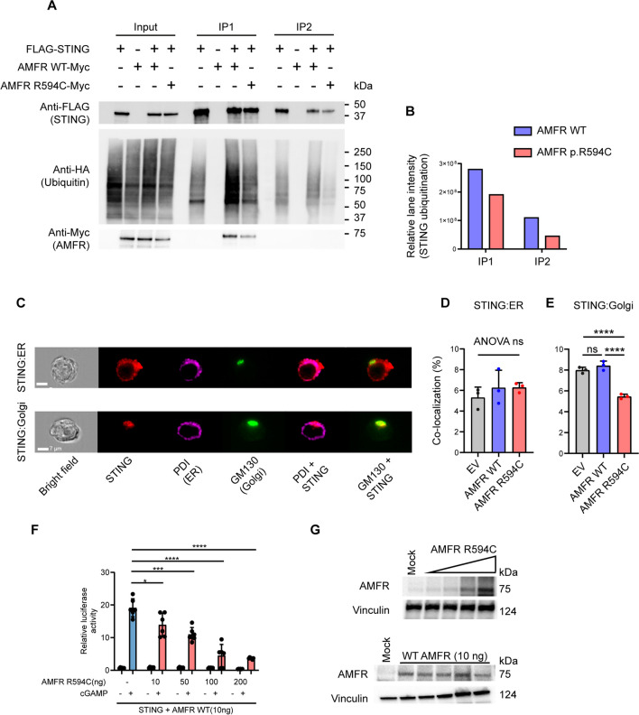
Fig. 6.
Decreased STING ubiquitination in cells expressing the patient AMFR R594C variant. A HEK293T cells were transfected with 1 µg of FLAG-STING, HA-K27 ubiquitin, and AMFR-Myc-WT or AMFR-Myc-R594C for 24 h, after which STING was immunoprecipitated twice with anti-FLAG-M2 beads. Lysates were immunoblotted for expression of FLAG-STING, HA-(K27) ubiquitin, and Myc-AMFR. B HA-ubiquitin expression was quantified in ImageLab (Bio-Rad, USA) by measuring the total lane intensity of the HA-ubiquitin smear for both AMFR-WT and AMFR-R594C mutant expressing cells in IP1 and IP2. The experiments are representative of three independent experiments. C–D HEK293T cells were transfected as indicated and probed with mouse anti-STING and anti-PDI (ER marker) or anti-GM130 (Golgi marker). Cells were analyzed by ImageStream. Panel C shows representative images, and panels D and E show quantification of STING-ER colocalization and STING-Golgi colocalization from three independent experiments of STING-ER co-localization F, G HEK 293T cells with stable STING expression were transfected with 10, 50, 100, 200 ng AMFR R594C or empty vector, IFNB1 promoter luciferase reporter, and β-actin Renilla reporter. F Eighteen hours after transfection, the cells were stimulated with 2′3′-cGAMP (50 mg/mL) for 8 h. G Cells were lysed for western blotting for AMFR and vinculin (loading control). Statistics were calculated with a 2-way ANOVA and Dunnett’s multiple comparisons test. *p<0.05; ***p < 0.001; ****p < 0.0001. The experiments were performed twice

