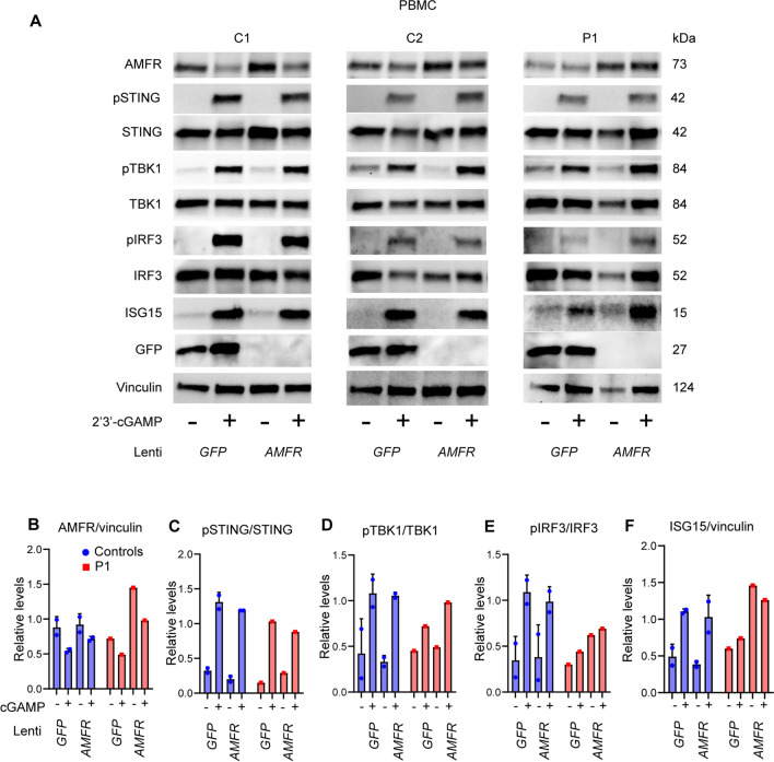
Fig. 7.
Reconstitution of patient PBMCs with WT AMFR restores cGAS-STING signaling and ISG responses. A Patient and control PBMCs were stimulated with 1.5 µg/mL PHA for 72 h and then transduced by VSV-G lentiviral vectors to express AMFR WT or GFP (control) (MOI 10). After 72 h, the cells were stimulated with 2′3′-cGAMP for 3 h, and cell lysates were analyzed by immunoblotting for AMFR, pSTING, STING, pTBK1, TBK1, pIRF3, IRF3, ISG15, GFP, and vinculin (loading control). (B–F) Quantification of the Western blot bands in (A)

