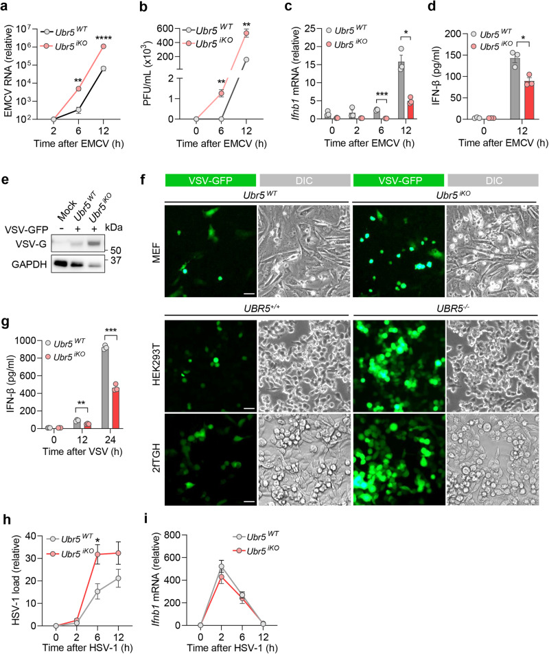
Fig. 3. UBR5 is crucial for the induction of type I IFNs by RNA viruses.
Quantification of (a) intracellular viral RNA by qRT-PCR, and (b) extracellular viral titers (plaque forming units, PFU/mL), in MEFs infected with EMCV at a multiplicity of infection (MOI) of 0.1. Quantification of (c) the cellular Ifnb1 mRNA by qRT-PCR and (d) secreted IFN-β by ELISA. e The immunoblot of cellular VSV glycoprotein (VSV-G) in MEFs. f Fluorescent images of VSV-GFP in three cell types. DIC: differential interference contrast. Scale bar: 50 µM. g Quantification of secreted IFN-β protein in MEFs, infected with VSV-GFP at a MOI of 0.5 for 24 h. Quantification of (h) intracellular HSV-1 RNA and (i) Ifnb1 mRNA by qRT-PCR, in MEFs infected with HSV-1 at a MOI of 0.5. Data shown in a–d, g–i are presented as mean ± S.E.M, two-tailed Student’s t test, n = 3 biologically independent experiments; for a: **p = 0.0038, ****p < 0.0001; for b: **p = 0.0040, **p = 0.0040 in sequence; for c: ***p = 0.0002, *p = 0.0133; for d: *p = 0.0297; for g: **p = 0.0075, ***p = 0.0002; for h: *p = 0.0299. Adjusted p values are presented. Source data are provided as a Source Data file.

