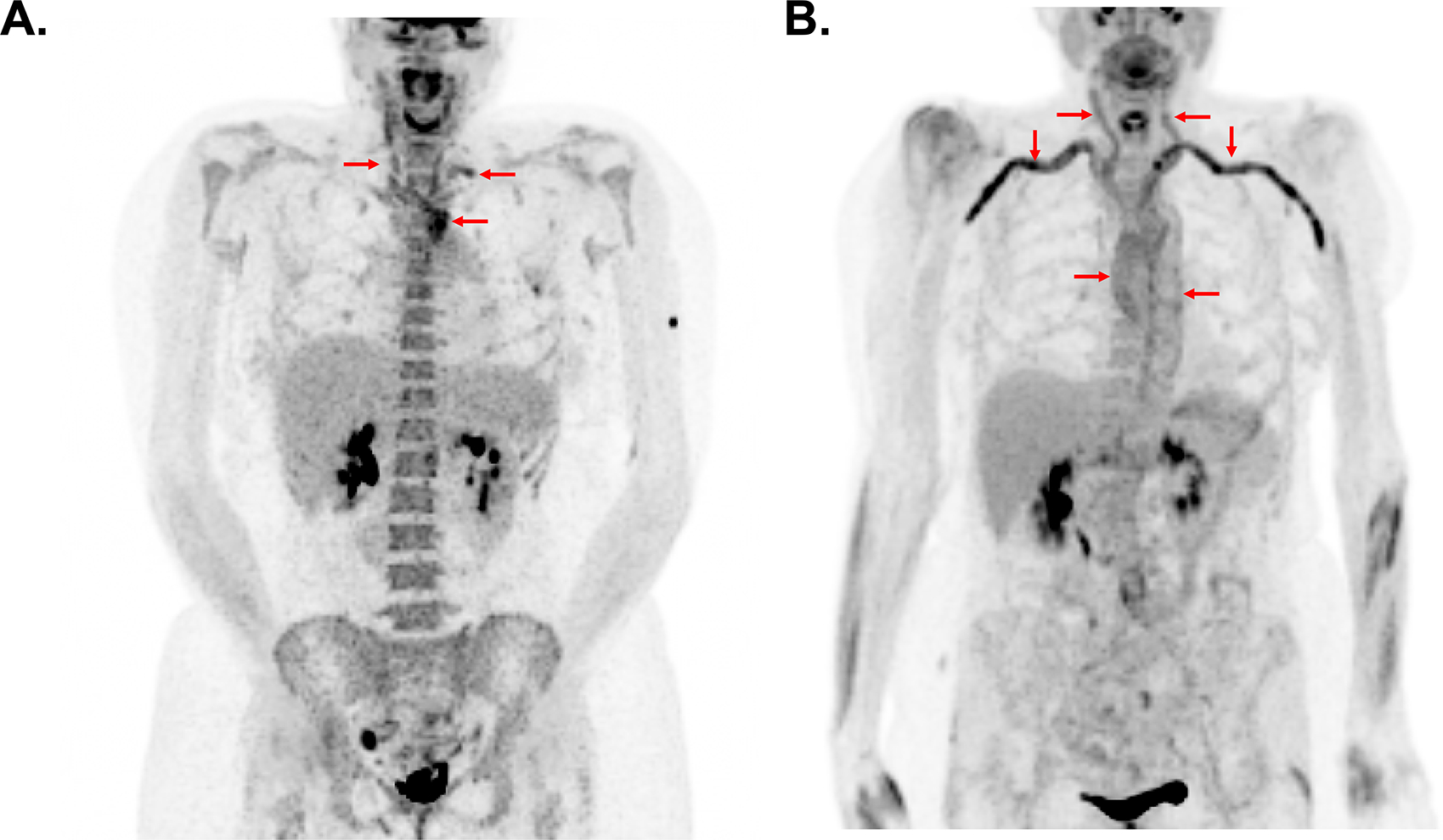
Figure 1. Typical Patterns of Arterial FDG Uptake in Takayasu’s Arteritis Compared to Giant Cell Arteritis.

While there is considerable variability in the pattern of vascular inflammation among patients with large-vessel vasculitis, patients with Takayasu’s arteritis commonly have focal areas of FDG uptake in the large arteries and patients with giant cell arteritis frequently have a more diffuse pattern of vascular FDG uptake. In panel A, a patient with Takayasu’s arteritis has intense FDG uptake confined to the aortic arch carotid arteries, and left subclavian artery (arrows). In panel B, a patient with giant cell arteritis has diffuse activity throughout the large arteries with particularly prominent signal in the subclavian and axillary arteries (arrows).
