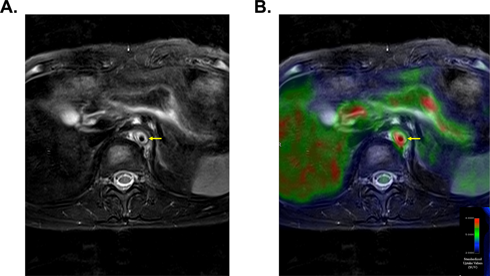
Figure 3. Multimodal Imaging with Angiography and FDG-PET Identifies High Risk Vascular Lesions.

Magnetic resonance imaging demonstrates vascular wall thickening and severe edema involving the descending aorta (Panel A). Concomitant FDG-PET-MR imaging demonstrates severe FDG uptake within the same vascular lesion (Panel B). This vascular area is at high risk for progressive vascular damage.
