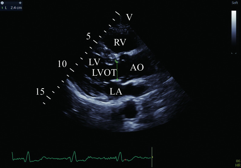
Figure 2.
Measurement of LVOTd.
A two-dimensional echocardiographic image was obtained using a GE Vivid E95 echo scanner equipped with a M5S electronic phased array probe (frequency 1.5–4.0 MHz). LVOTd was determined based on the parasternal long-axis view when the systolic aortic valve was fully opened. Abbreviations: LVOTd, left ventricular outflow tract diameter; LVOT, left ventricular outflow tract; LA, left atrium; LV, left ventricle; RV, right ventricular; AO, aorta.

