FIG. 7.
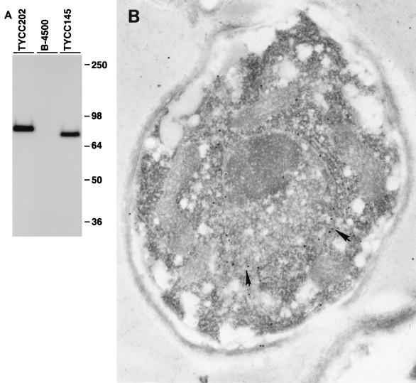
Localization of Cap60p. (A) Immunoblot analysis. Yeast cells were grown in YNB, and total protein extracts were analyzed by sodium dodecyl sulfate–8% polyacrylamide gel electrophoresis and incubated with anti-HA antibody and the Western-Star chemiluminescent detection system. B-4500 is a wild-type strain; TYCC145 is an encapsulated transformant containing a plasmid with a single HA-tagged CAP60; and TYCC202 is an encapsulated transformant containing a plasmid with a three-HA-tagged CAP60. (B) Electron micrograph of TYCC202. The yeast cells grown in YNB were fixed in equal volumes of 8% formaldehyde and 0.2% glutaraldehyde. Ultrathin sections (50 to 60 nm) were incubated with anti-HA serum and with 15-nm-diameter colloidal gold-conjugated goat anti-mouse IgG secondary antibody. The thin sections were counterstained with uranyl acetate and lead citrate. The antibodies are associated with the nuclear envelope (arrows). The picture shown is representative of many sections. Magnification, ×3,400.

