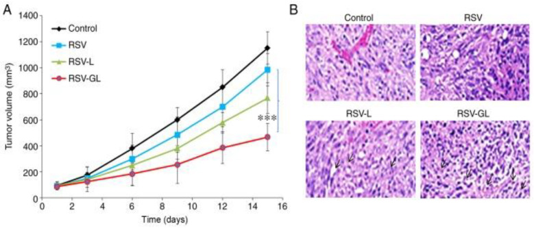
Figure 6.
In vivo analysis of RSV, RSV-L, and RSV-GL in SSC-bearing xenograft model. (A) Tumor volume; (B) hematoxylin and eosin histology staining analysis. *** p < 0.0001 vs. RSV. Reprinted from Ref. [141]. Copyright 2019, International Journal of Molecular Medicine. This work is licensed under Attribution-NonCommercial-No Derivatives 4.0 International (CC BY-NC-ND 4.0).

