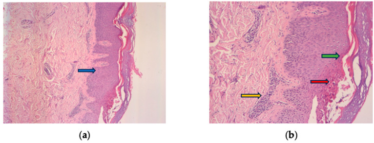
Figure 2.
Histologic findings typically of psoriasis: epidermal hyperplasia (blue arrow), parakeratosis (green arrow), Munro’s micro abscesses (red arrow), thinned granular cell layer of the epidermis, dilated dermal capillaries, and inflammatory infiltrate in the upper dermis (yellow arrow). (a) HE 10× and (b) HE 30× magnification.

