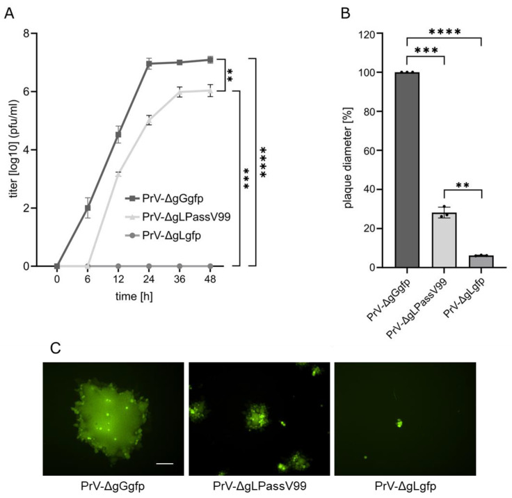
Figure 1.
Replication properties of PrV-ΔgLPassV99. (A) RK13 cells were infected with PrV-ΔgGgfp, PrV-ΔgLPassV99 and PrV-ΔgLgfp at a MOI of 0.5. Cells and supernatant were harvested at the indicated times after infection and titers were determined on RK13 cells. Mean titers in log10 plaque forming units [pfu]/mL and corresponding standard deviations are given. (B) RK13 cells were infected under plaque assay conditions and plaque diameters were measured two days post infection. Shown are mean percent values compared to PrV-ΔgGgfp plaques set as 100% and corresponding standard deviation from three different experiments. Two-tailed Welch’s t test; **, p < 0.01; ***, p < 0.001; ****, p < 0.0001. (C) Representative images of plaques formed one day post infection. Scale bar: 100 µm.

