Figure 2.
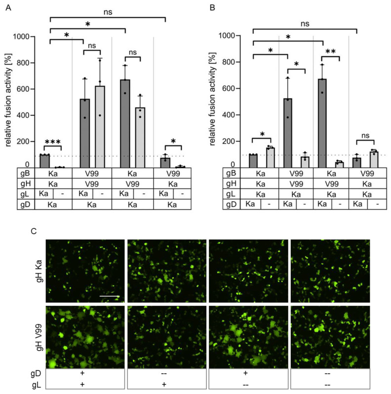
In vitro fusion assays. (A) RK13 cells were transfected with expression plasmids encoding gB and gH derived either from PrV-Ka (Ka) or PrV-ΔgLPassV99 (V99), gD Ka and with or without (-) pcDNA-gL. (B) Plasmids encoding either wild type gL, gB, gH or gB and gH derived from PrV-ΔgLPassV99 were cotransfected into RK13 cells either with or without (-) pcDNA-gD Ka. Area and number of syncytia was measured 19 h post transfection and calculated corresponding to assays with plasmids expressing Ka gB, gD, gH and gL set as 100%. Shown are mean values of three independent assays with corresponding standard deviations. Two-tailed Welch’s t test; ns, not significant; *, p < 0.05; **, p < 0.01; ***, p < 0.001. Values for panels A and B were measured in combined assays but separated for clarity. (C) Representative images of syncytia formed after cotransfection of RK13 cells with gB Ka and gH Ka or gH V99 in presence (+) or absence (--) of gD and gL. Scale bar: 200 µm.

