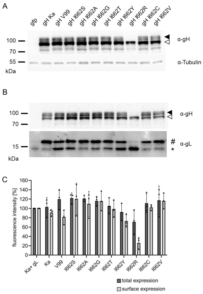
Figure 4.
Expression and processing of the different gH variants. (A) RK13 cells were transfected with expression plasmids for gH Ka or the different gH mutants and cell lysates were harvested one day post transfection. After Western blotting, the membrane was cut between the 70 and 55 kDa markers and the upper part was probed with a monospecific rabbit α-gH serum and the lower part with an anti-tubulin monoclonal antibody as loading control. (B) RK13 cells were cotransfected with expression plasmids for gL and the different gH variants. Parallel blots were probed with anti-gH and anti-gL sera. Immature and mature forms of gH are indicated by open and filled arrow heads, and of gL by an asterisk (*) and a diamond (#), respectively. Cells transfected with pEGFP-N1 were used as negative control. Molecular masses of marker proteins in kDa are given. (C) Relative total or cell surface fluorescence intensities of cells transfected with the indicated expression constructs are given. Cells cotransfected with pcDNA-gH Ka and pcDNA-gL were set as 100%, and cells transfected with the empty vector were used as background control. Mean values of three independent assays and standard deviations are shown.

