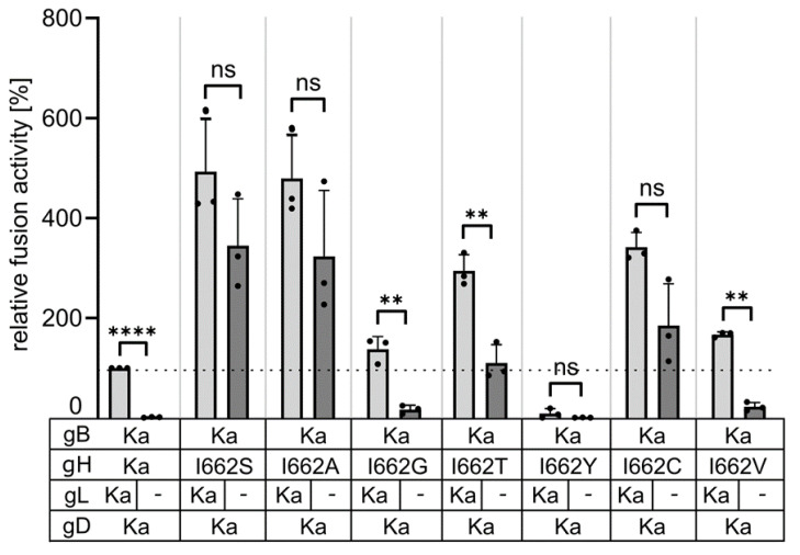
Figure 5.
Different amino acids at position 662 in the gH TMD compensate for lack of gL in cell–cell fusion assays. Expression plasmids encoding gD Ka, gB Ka, gH Ka or mutated gH as indicated were cotransfected either in presence (black bars) or absence (grey bars) of pcDNA-gL into RK13 cells. Relative fusion activity was determined 19 h post transfection. Mean values of three independent assays and corresponding standard deviation are given. Two-tailed Welch’s t test; ns, not significant; **, p < 0.01; ****, p < 0.0001.

