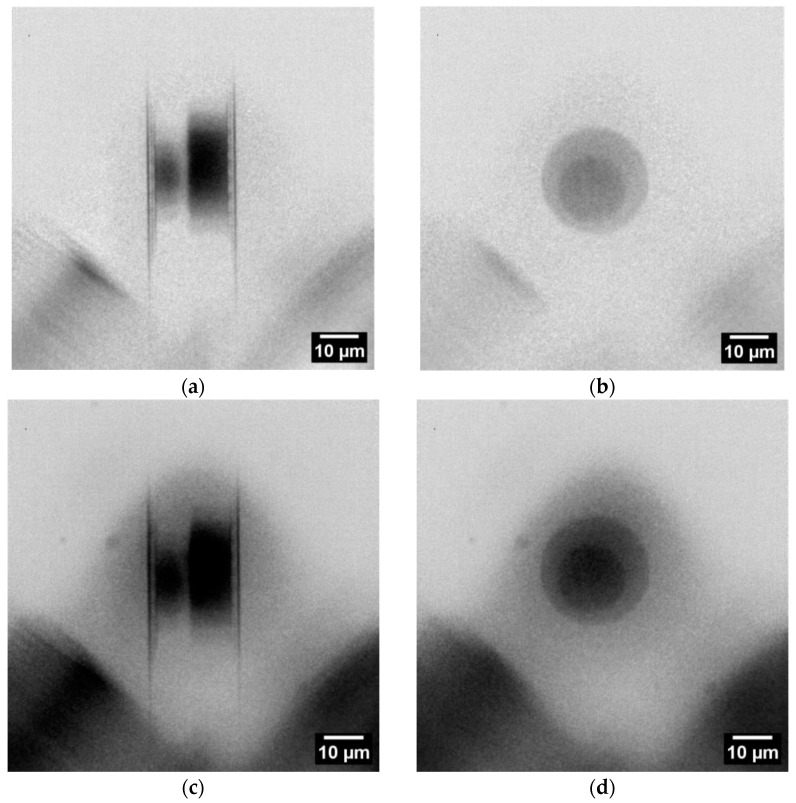
Figure 4.
Radiographs of the SLID sample (rotated left by straight angle compared to Figure 3b) in two imaging directions: (a) at 90° cross-section view with visible solder of top and bottom dies and, in between, a light gray layer of IMC; (b) at 0° the plane view (stacking direction). (c,d) are similar to (a,b) with the addition of a 680 µm Si wafer piece in the beam path. Each radiograph is the result of five single radiographs with exposure time of 120 s using the camera binning 2 mode (1024 px × 1024 px).

