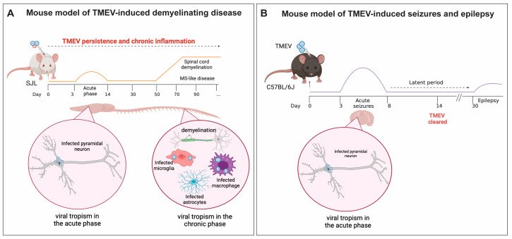
Figure 1.
Schematic representation of (A) Mouse model of Theiler’s murine encephalomyelitis virus-induced demyelination disease (TMEV-IDD): Intracerebral (IC) infection of SJL/J mice with TMEV results in a persistent viral infection throughout the mouse’s lifespan. This is a biphasic disease; an acute phase occurs between 3 and 14 dpi; chronic neuroinflammation due to viral persistence in the central nervous system (CNS) leads to a multiple sclerosis (MS)-like phase, which appears 40–60 days post-infection. In this phase, weakness of the hind limbs and ataxic paralysis are observed. During the acute phase, TMEV primarily infects CA1 and CA2 hippocampal neurons and leads to the activation of innate and adaptive immune responses. Due to failure in viral clearance, chronic neuroinflammation is observed during MS-like disease and is characterized by viral persistence in the white matter of the spinal cord, and oligodendrocytes (not shown here), astrocytes, microglia, and infiltrating macrophages are the main cells infected by TMEV during this phase. (B) Mouse model of viral-induced seizures and epilepsy. C57BL/6J mice ICinfected with TMEV develop encephalitis. Between 3 and 8 dpi, mice develop acute seizures, followed by a latent period in which seizures are no longer observed. Between 30 and 100 dpi, a portion of the mice that experienced acute seizures develop spontaneous recurrent seizures (epilepsy). During the acute phase of the infection, TMEV infects pyramidal neurons in the CA1 and CA2 regions of the hippocampus. Induction of innate and adaptive immune response results in viral clearance, but also contributes to neuronal excitation and seizure development. Figure made using Biorender.

