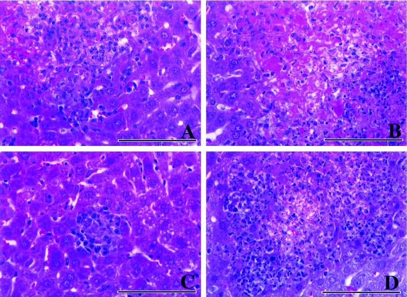
FIG. 2.
Hematoxylin-and-eosin staining of liver sections from infected mice 4 days after inoculation with an immunizing dose of L. monocytogenes. (A) Control mouse. Macrophages predominate, with lymphocytes and scattered neutrophils also present. (B) αβ-KO mouse. A large area of severe hepatic necrosis is evident, with a rim of degenerate neutrophils present (right side). (C) γδ-KO mouse. A small focus of infection can be seen in this section, which contains lymphocytes and macrophages. (D) IFN-KO mouse. An area of necrosis is surrounded by a large number of inflammatory cells, most of which are neutrophils. Bars, 100 μm.

