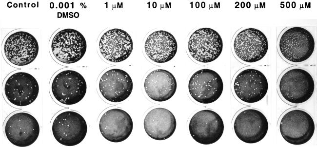
FIG. 9.
Influence of α-lipoic acid on the appearance of R. rickettsii plaques in Vero cells. One milliliter of each 10−5 to 10−8 10-fold serial dilution of a standard seed of R. rickettsii was inoculated, in triplicate, in 22-mm-diameter wells, with a subsequent 1-h incubation at 35°C. The inocula were aspirated, and the infected monolayers were overlaid with 0.5% agarose prepared in cell culture medium with or without α-lipoic acid supplementation. Plaques were counted on day 7 after inoculation, following staining with 0.3% neutral red. From the top: first row, 10−6 dilution; second row, 10−7 dilution; third row, 10−8 dilution.

