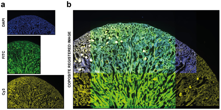
Figure 4.
Automatic registration. Example of usage of DS4H-IA. (a) Input images referring to a commercial mouse kidney biopsy (FluoCellTM prepared slide #3, Invitrogen). From top to bottom: Fluorescence DAPI (nuclear staining, D-1306), FITC (cytoplasmic staining, Alexa Fluor 488 wheat germ agglutinin), and Cy3 (membrane staining, Alexa Fluor 568 phalloidin) images all acquired using a Nikon A1R confocal Microscope equipped with a 20x objective. (b) Example of a common output registered stack. In this example, just the DAPI, FITC, and Cy5 signals are shown.

