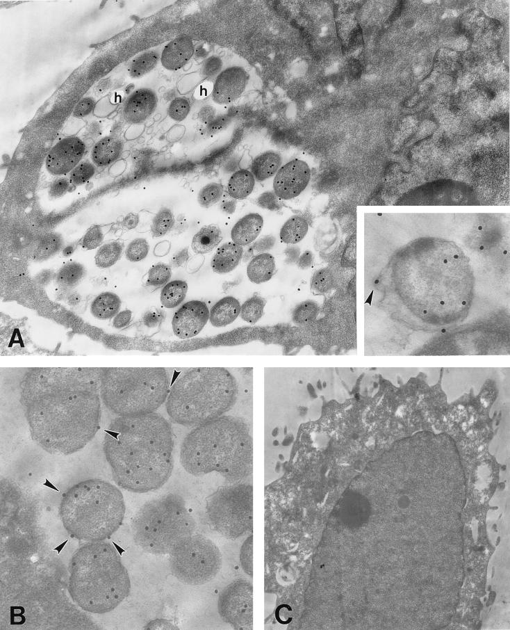
FIG. 1.
Localization of chlamydial hsp60 in infected HEC-1B cells. Thin sections were probed with a chlamydia-specific hsp60 monoclonal antibody and labeled with 30-nm-diameter gold-conjugated second-affinity antibodies. Labeling of hsp60 in normally infected cells (A and B) at 48 h p.i. and in uninfected cells (control) (C) is shown. The arrowheads indicate gold particles associated with the chlamydial cell envelope (A, enlarged inset, and B). Translucent holes (h) are often observed in samples embedded in the fragile Lowicryl resin. Magnifications: ×15,750 (A); ×35,777 (inset); ×16,285 (B); and ×7,700 (C).

