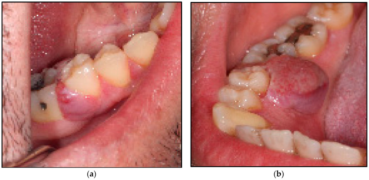
Figure 2.
Shows the clinical presentation of the oral mucosal lesions. (a) represents erythromatous, pedunculated lesion with irregular texture, moderate in consistency, about 1 × 1 cm in diameter occupying the buccal gingivae and the entire interdental region between LR5 and LR6 buccally, firmly attached to the underlining structures; (b) represents a lobulated erythromatous, spongy (moderate in consistency) lesion about 1 × 1.5 × 2 cm in size, occupying the entire interdental papillae and the lingual mucosa between LR5 and LR6, firmly attached to the underlining structures.

