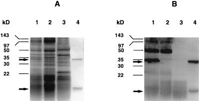
FIG. 3.
Proteins from supernatants of mitomycin C cultures and SLT-II holotoxin were separated on a sodium dodecyl sulfate-polyacrylamide gel (A) and exposed to a polyclonal pig anti-SLT-II serum in the corresponding Western blot (B). EHEC strain 86-24 is shown in lanes 1, E. coli TUV86-2 is shown in lanes 2, E. coli C600 is used as a negative control in lanes 3, and SLT-II holotoxin is used as a positive control in lanes 4. Molecular masses were estimated by utilizing a prestained marker from Bio-Rad. Arrows indicate the position of the SLT-II A- and B-subunits at 32 and 7 kDa, respectively.

