Fig. 1.
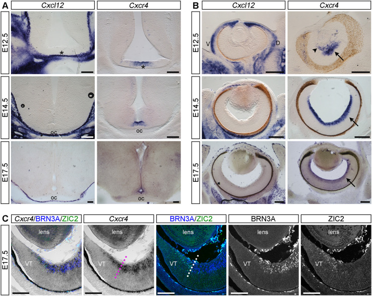
Cxcl12 is expressed by ventral diencephalon meninges and Cxcr4 is expressed by RGCs. (A,B) In situ hybridisation for Cxcl12 and Cxcr4 on coronal vibratome sections through the ventral diencephalon (A) and retina (B) of E12.5, E14.5 and E17.5 mouse embryos. Asterisks in A indicate the position in the ventral diencephalon where the optic chiasm (oc) will form. In B, arrows indicate expression of Cxcr4 in the RGC layer of the retina and arrowhead indicates the hyaloid vasculature. (C) Cryosection through the E17.5 peripheral ventrotemporal (VT) retina stained after Cxcr4 in situ hybridisation by immunofluorescence with antibodies specific for BRN3A (labels contralaterally projecting RGCs) and ZIC2 (labels ipsilaterally projecting RGCs). Staining is shown as the combined and single channels. Dotted lines indicate the boundary between ZIC2-positive and -negative RGCs. Scale bars: 200 µm (A,B); 100 µm (C). D, dorsal; V, ventral; VT, ventrotemporal.

