Fig. 2.

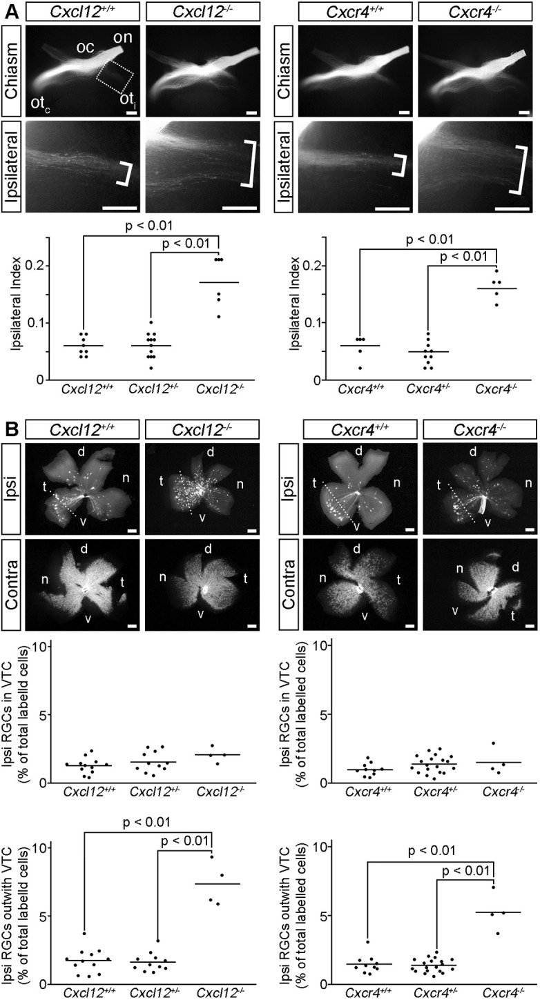
CXCL12 and CXCR4 are required for contralateral RGC axon growth at the optic chiasm. (A) Ventral views of RGC axons labelled from one eye in E14.5 Cxcl12 and Cxcr4 wild-type and mutant littermates captured using a stereo microscope. The optic nerve (on), optic chiasm (oc), contralateral optic tract (otc) and ipsilateral optic tract (oti) are indicated. Boxed region is shown at higher magnification in the lower panels; brackets indicate the width of the ipsilateral projection. Number analysed of each genotype was: Cxcl12+/+ n=8, Cxcl12+/− n=12, Cxcl12−/− n=6; Cxcr4+/+ n=5, Cxcr4+/− n=10, Cxcr4−/− n=5. (B) Ipsilateral (Ipsi) and contralateral (Contra) flatmounted retinas from retrogradely labelled E15.5 Cxcl12 and Cxcr4 wild-type and mutant littermates. Dotted line demarcates the ventrotemporal crescent (VTC). d, dorsal; n, nasal; t, temporal; v, ventral. Number analysed of each genotype was: Cxcl12+/+ n=12, Cxcl12+/− n=11, Cxcl12−/− n=4; Cxcr4+/+ n=10, Cxcr4+/− n=19, Cxcr4−/− n=4. Graphs display quantification of the Ipsilateral Index (A) and proportion of ipsilaterally projecting RGCs relative to the total number of labelled cells in both eyes (B), with each dot representing the value for one embryo and horizontal bars the mean values. Statistical analyses were performed using one-way ANOVA with Tukey post-hoc comparison. Scale bars: 200 µm.
