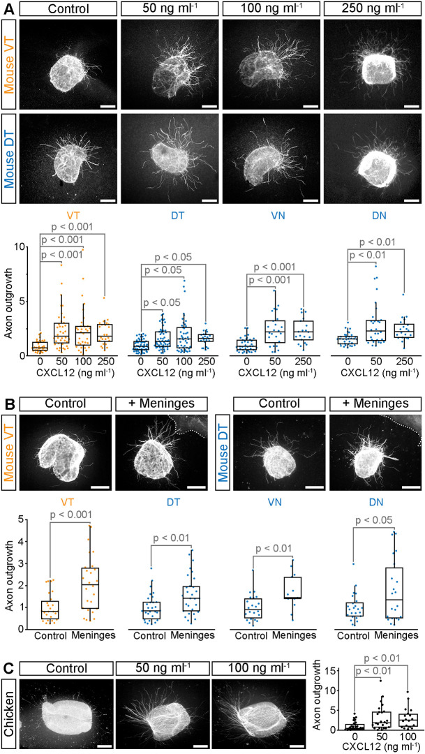
Fig. 4.
CXCL12 and ventral diencephalon meninges promote RGC axon outgrowth. (A-C) Representative examples and quantification of E14.5 mouse (A,B) or E6 chicken (C) retinal explants cultured in the presence and absence of CXCL12 (A,C) or ventral diencephalon meninges (B). Mouse explants from peripheral ventrotemporal (VT) retina contain predominately ipsilaterally projecting RGCs; peripheral dorsotemporal (DT), ventronasal (VN) and dorsonasal (DN) explants contain contralaterally projecting RGCs. All RGCs in chicken project contralaterally. Dotted lines in B indicate the edge of the meningeal tissue. Quantitative data are shown as boxplots with the median values (middle bars) and first to third interquartile ranges (boxes); whiskers indicate 1.5× the interquartile ranges; each dot represents the value from one explant. A minimum of 22 explants from three independent experiments (A), 13 explants from three independent experiments (B) or 18 explants from four independent experiments (C) were analysed per condition. Statistical analyses were performed using Kruskal–Wallis rank sum test with TUKEY-Kramer post-hoc comparison. Scale bars: 200 µm.

