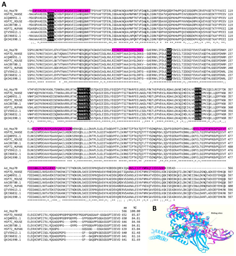
Figure 9.
Hsp 70 kDa. (A) Multiple alignments of the amino acid sequence of putative A. dowii Hsp 70 (Ad_Hsp70 (Transcriptome: c27026_g1_i1)) with other proteins of the same family. In the alignment, the sequences Hsp70 from insects (UniProtKB: HSP7D_MANSE), from humans (UniProtKB: HSP72_HUMAN), from mice (UniProtKB: HSP7C_MOUSE), and several patented Hsp70s for different applications (GenBank: ACQ4089780.1, A QTV59613.1, QRJ96032.1, QKO41990.1). The region covered by the tryptic peptides obtained from the proteomes of A. dowii and L. neglecta is highlighted by a magenta background. The ATP binding site is underlined and the regions involved in the binding of nucleotide phosphates are highlighted with white letters on a black background. (B) Structure models of heat shock proteins: Ad_Hsp70 is green, HSP7D_MANSE is medium blue, ACQ40951.1 is blue, HSP7C_MOUSE is forest green, AAC89780.1 is gray, HSP72_HUMAN is pink, QTV59613.1 is yellow, QRJ96032.1 is red, and QKO41990.1 is purple. The comparison of the Cα chains presented RMSD values that oscillate between 0.007 and 0.77.

