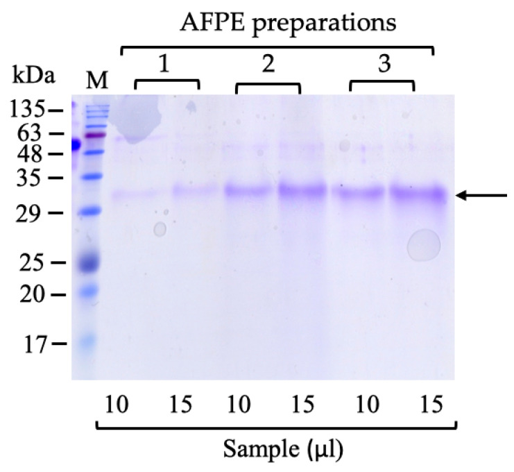
Figure 1.
Protein analysis of AFPE by SDS-PAGE: Three different AFPE preparations (1, 2, and 3) were loaded (10 and 15 μL) onto SDS-PAGE, which was carried out using 15% polyacrylamide separating gel, and stained with Coomassie R-250 blue. M, molecular weight marker; the arrow shows the ~33 kDa protein.

