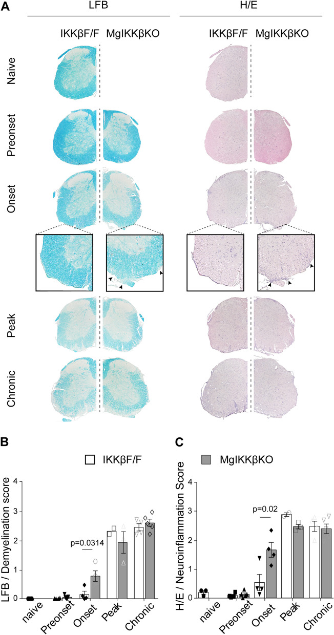
Fig. 4.
Knocking out IKKβ selectively from CNS macrophages results in early demyelination and neuroinflammation in EAE. A Specimen images of paraffin-embedded coronal spinal cord sections from IKKβF/F and ΜgΙΚΚβKO mice stained with LFB (left column) and H/E (right column) showing the evolution of demyelination and immune cell infiltration, respectively, during MOG35-55-induced EAE. Specifically, in the naïve spinal cord, at a pre-onset stage (dpi 8), at the onset (of the ΜgΙΚΚβKO mice), at the peak and at the chronic phase of the disease. B Semi-quantification of the mean demyelination level in spinal cord of IKKβF/F and ΜgΙΚΚβKO mice shown in A, left column. C Semi-quantification of the mean immune cell infiltration level in spinal cord of IKKβF/F and ΜgΙΚΚβKO mice shown in A, right column. Numbers of mice are annotated as scatter dots on the bars. All mice were adult females 2–4 months old

