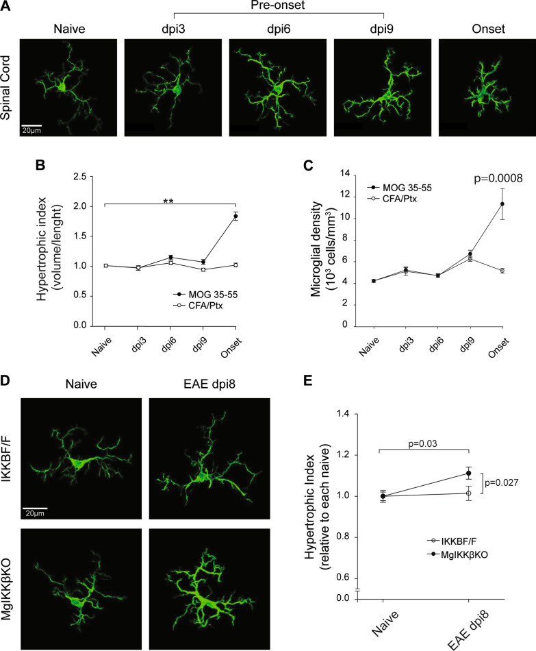
Fig. 5.
Spinal cord parenchymal microglia change morphology at the pre-onset stage of EAE in ΜgΙΚΚβKO mice. A Representative confocal images of individual microglia from cryostat slices immunolabeled for Iba-1 showing the spatiotemporal evolution of microglial morphology at the spinal cord of female C57BL/6 mice during MOG35-55-induced EAE. Specifically in the naïve spinal cord, at a pre-onset stage (dpi 3, 6 and 9) and at the onset of the disease. B Quantification of the microglial relative hypertrophic index (volume/length) in the spinal cord of C57BL/6 mice mice with MOG35-55-induced EAE or in controls that had been injected with the CFA adjuvant only and the Pertussis toxin (CFA/Ptx). C Quantification of microglial density in the spinal cord of C57BL/6 mice mice with MOG35-55-induced EAE or in controls that had been injected with CFA adjuvant only and Pertussis toxin (CFA/Ptx). D Representative confocal images of individual microglia from cryostat spinal cord slices from IKKβF/F and ΜgΙΚΚβKO mice that were either naïve or at the dpi 8 pre-onset stage of MOG35-55-induced EAE. E Quantification of the microglial relative hypertrophic index in the spinal cord of naïve and EAE dpi8 IKKβF/F and ΜgΙΚΚβKO mice. All mice were adult females 2–4 months old

