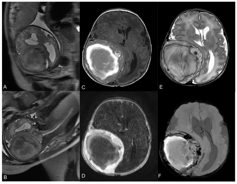
Figure 1.
Fetal MR at 35-week gestational age—T2-w axial (A) and coronal (B) images show a large heterogeneous lesion (69 × 62 × 67 mm3) with severe mass effect on the surrounding brain. Neonatal pre-operative head MR performed 2 days after birth. Axial T1-w image (C) shows the hyperintense hemorrhagic component. Axial T2-w (E) demonstrates the mass effect on the right parietal lobe, with right lateral ventricle compression and contralateral midline shift (15 mm). Axial T1-w image after Gadolinium administration (D) demonstrates a peripheral solid part with intense enhancement. Axial SWI sequence (F) shows multiple hypointense spots within the lesion, indicative of hemosiderin.

