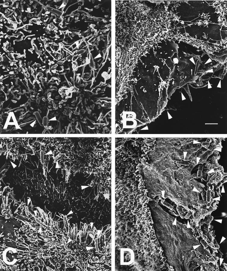
FIG. 4.
High-resolution SEMs of bacteria (highlighted by white arrowheads) adherent to enterocytes preincubated for 1 h in medium supplemented with 1 μg of cytochalasin D per ml. (A) L. monocytogenes diffusely adherent to the apical surface of a HT-29 enterocyte, showing listerial cells entwined among the apical microvilli; (B) P. mirabilis preferentially adherent to the lateral surface of a Caco-2 enterocyte with its apical surface devoid of bacteria; (C and D) S. typhimurium preferentially adherent to the lateral surfaces of two separated HT-29 enterocytes (C) as well as to the lateral surface of a Caco-2 enterocyte (D), where bacteria appear both tightly adherent to the lateral surface as well as in the process of internalization into the Caco-2 cytoplasm. Scale bars: A, 1 μm; B, 2.5 μm; C, 2.2 μm; and D, 1.7 μm.

